3D Microcosm of Lichens: Researchers Visualize Fine Structure of Lichen Symbiosis
Botany, 16.09.2026
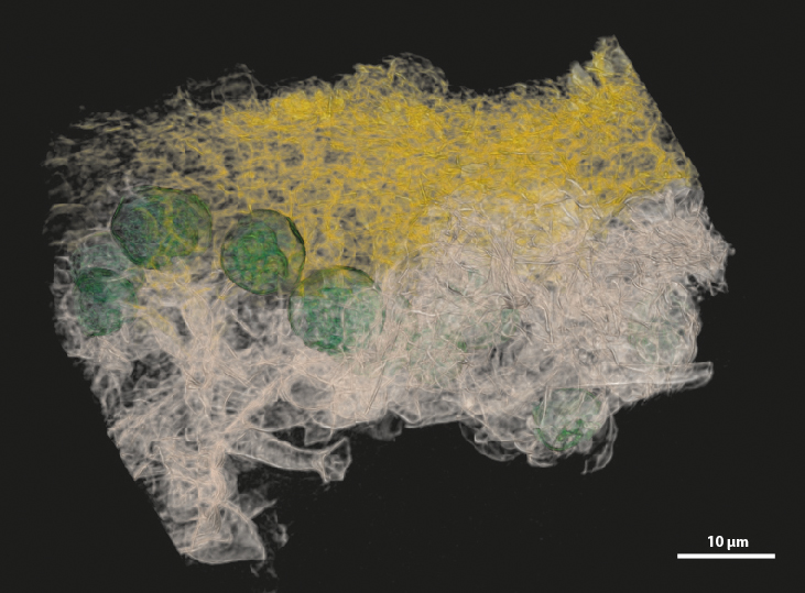
Lichens are symbiotic communities of fungi and algae. Researchers led by SNSB botanist PD Dr. Andreas Beck have now, for the first time, visualized in 3D the areas within the lichen thallus where the cells of the two symbiotic partners come into contact and exchange substances. The cell walls at the fungus-algae contact zone show distinct changes in their structure and chemical composition. The researchers have now published their findings in the journal Scientific Reports.
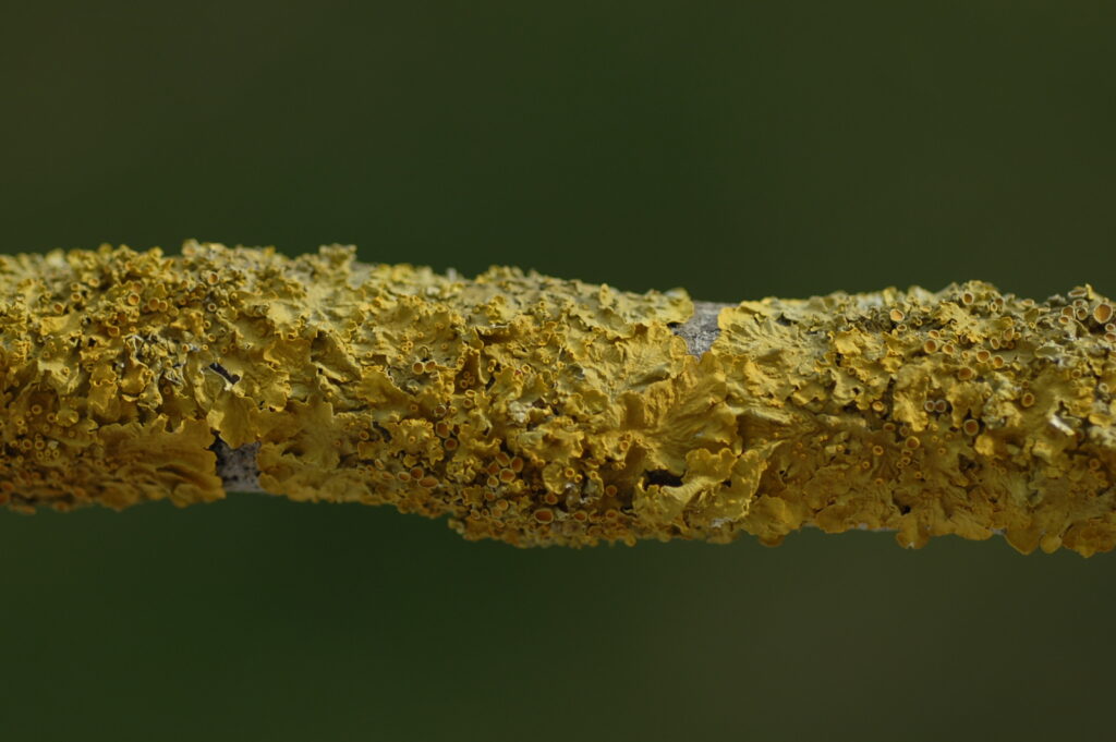
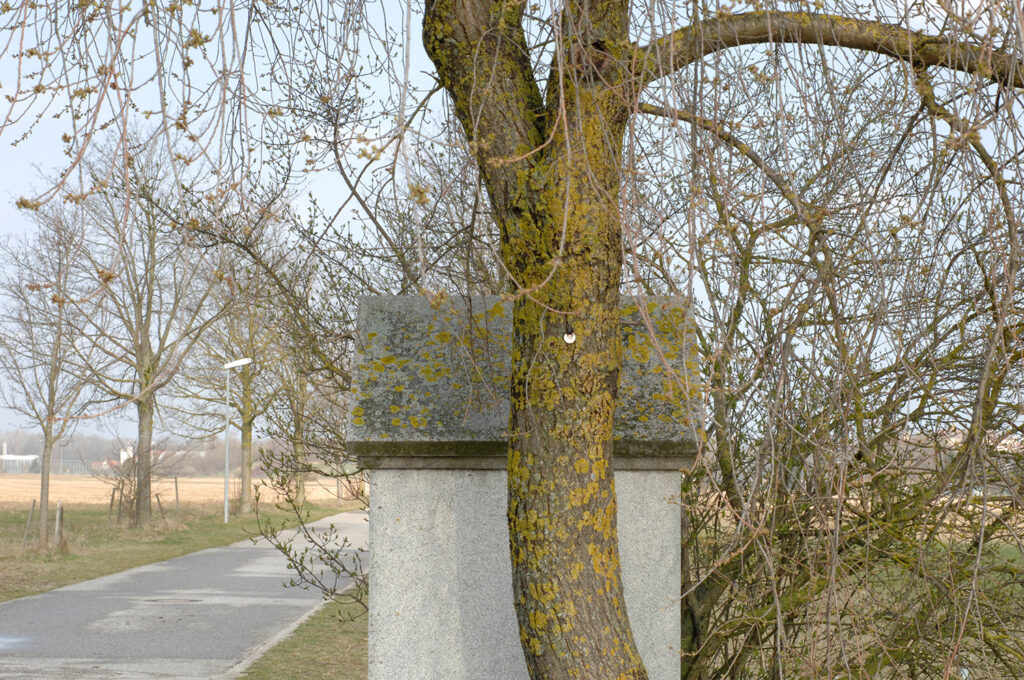
Lichens colonize trees or rocks all over the world. They are symbiotic communities between fungi and photosynthetically active green algae or cyanobacteria, in which both partners benefit from each other: The fungal partner relies on the algae for a supply of carbohydrates. In return, it protects the algae from UV radiation and other environmental influences. The contact zone where the fungal cells and algal cells touch is of crucial importance for the transfer of nutrients between the two symbiotic partners. Detailed investigations, particularly a three-dimensional understanding of this area between the cells, have been lacking until now.
A research team led by botanist PD Dr. Andreas Beck of the Bavarian State Collections of Natural History (SNSB), in collaboration with Prof. Dr. Christian Bayer of Aalen University of Applied Sciences, has now succeeded for the first time in visualizing this interaction zone between the two symbiotic partners of the wall lichen Xanthoria parietina in three dimensions. The high-resolution nano-CT images show in detail where and how the fungus and algae come into contact via their cell walls—areas inside the lichen that are only a few ten-thousandths of a millimeter in size. For comparison: an average human hair is about 200 times thicker than these structures. It becomes apparent that the algae are directly connected to the filamentous fungal cells via about one-quarter of their cell wall surface area. The contact area is further increased by a volume expansion of those fungal cells located in the immediate vicinity of the algal cells. Specialized material analyses, known as energy-dispersive X-ray spectroscopy, also show that the chemical composition of the cell walls at the fungus-alga contact zones has changed. Both the nitrogen and sulfur content are significantly elevated there, which the researchers suspect may reflect an influence on the transport processes between the symbionts.
“Our analyses show, for the first time, the precise modification of the symbiont cell wall in the contact area between the alga and the fungus. Genomic analyses have not provided us with these details so far. The 3D models of the interaction zone between the two lichen symbionts are of great interest for understanding their interaction. “Our findings contribute to a better understanding of the symbiotic metabolism of lichens and its potential applications,” explains first author Andreas Beck, discussing the study’s results.
Researchers from the Bavarian State Collections of Natural History, Aalen University of Applied Sciences and the Helmholtz Institute Freiberg participated in the study.
Publication:
Beck, A., Bayer, C., Debastiani, R., Schubert, T., Gröger, A., Ruthensteiner, B. (2026) Elemental composition, structure and amount of contact between Xanthoria parietina symbionts. Sci Rep 16, 27379 https://doi.org/10.1038/s41598-026-68076-7
Scientific Contact
PD Dr. Andreas Beck
SNSB – Botanische Staatssammlung München
Tel.: 089 17861 266, E-Mail: beck@snsb.de